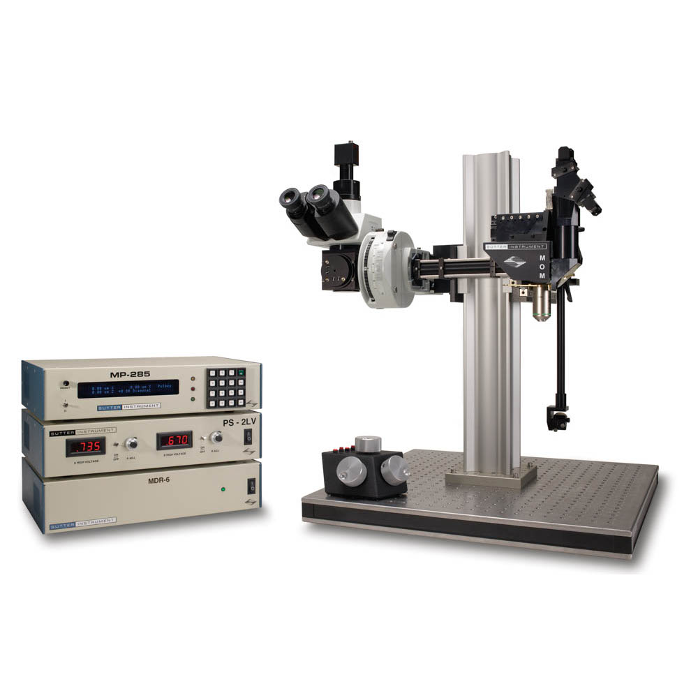
MOM Movable Objective Microscope
The Movable Objective Microscope® (MOM®) is a two-photon microscope capable of imaging deep within living specimens when combined with a Ti:Sapphire Laser. The MOM design is unique in providing 3-dimensional objective movement and rotation allowing the specimen to remain stationary.
Product Enquiry
If you would like to send us an enquiry about this product, please click the button below, fill in the form and submit.
Product Enquiry
Product Enquiry
Please fill out the form if you would like to enquire about this product."*" indicates required fields
The Movable Objective Microscope® (MOM®) is a two-photon microscope capable of imaging deep within living specimens when combined with a Ti:Sapphire Laser. The MOM design is unique in providing 3-dimensional objective movement and rotation allowing the specimen to remain stationary. Many highly regarded imaging laboratories around the world use the Sutter MOM and we constantly work with our customers to adapt the design for their changing needs.
Watch the video describing MOM imaging and photostimulation beam paths
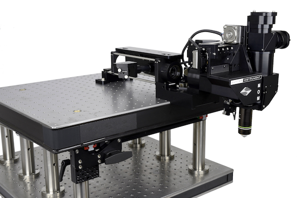
MOM Opto-mechanical Design
The MOM consists of two independent microscopes. The wide-field half of the microscope consists of an Olympus vertical illuminator, Sutter Xenon arc lamp and camera mount to provide standard epifluorescence. The two-photon side of the microscope provides the optical pathway for guiding the excitation laser light from the table up into the scanning galvanometric mirrors and then expanding the beam through the scan lens and directing into the back of the objective. Following two-photon excitation, the emitted photons are directed by a dichroic mirror immediately above the objective into the detection pathway. The main body of the microscope moves backwards on a rail system allowing easy access to the specimen prior to imaging.
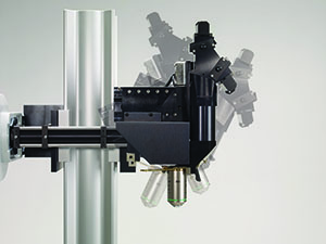
The objective translates in X, Y and Z as well as rotates around the X axis. Two moving mirrors allow the microscope to maintain efficient delivery of the excitation light to the back aperture of the objective regardless of movement or orientation. The X, Y and Z movements used are the same as that in our MP-285 micromanipulator so you know the movements are smooth, fine in scale, drift-free and highly reproducible. These movements permit Z-stacks and mosaic images of large regions of tissue to be recorded without the need for a moving stage.
The horizontal light path allows for rotation of the objective away from the standard vertical position. As a result of this rotation, the MOM can easily be converted from an upright to an inverted microscope and the objective positioned from 0 to 180 degrees. This positional freedom permits the imaging of non-horizontal surfaces and volumes.
MOM Scanning Systems
During the last 10 years, scanning technology has changed dramatically: objectives changed requiring larger beam sizes and scanner technology advanced providing dependable resonant scanners. Unlike other two photon microscope designs, the MOM has lived through and adapted to changes in scanning technology. Throughout this development, Sutter has maintained two principles. First, existing scopes can be upgraded to new technology as it becomes available. Many original scopes with 3mm galvo scanners have been upgraded to either 6mm galvo scanners or resonant/galvo scanners. Second, Sutter continues to supply the original designs if they are needed for current research. We can provide a 3mm or 6mm conventional scanning MOM or a resonant/galvo scanning MOM all at competitive prices.
Imaging Software
Starting in 2011, Sutter began offering the MOM Computer System and Software (MCS). Before this software package was developed, most users relied on ScanImage or MPScope to generate scanned images. Customers valued the fact that the MOM would operate with open source freewares, however, there seemed to also be a market for a commercial package. Sutter MScan offered a number of functions that were not available in the existing freeware packages including photostimulation and the ability to combine imaging with electrophysiological recording and photostimulation. As resonant scanning became popular, and none of the freewares supported resonant scanning on the MOM, Sutter and MScan took the initiative. The latest version, MScan 2.0, coupled with a faster data acquisition system, allowed the MOM to generate fast, resonant images. To this day, the Sutter MOM with MScan2.0 remains the one platform available that can switch back and forth between conventional and resonant scanning.
The MOM® has always been compatible with ScanImage freeware, the two-photon imaging software developed by Karel Svoboda and collaborators. One of the reasons the MOM platform exists in its present form is the strong support from the ScanImage community. In 2014, Vidrio became the principle vehicle for support and new development of ScanImage. Sutter is happy to make Vidrio ScanImage Premium available to customers who wish premium support and the latest features. ScanImage freeware is also still available as SI5. Sutter provides packages that include the necessary data acquisition hardware to couple the MOM and other scanning microscopes to ScanImage Premium or SI5.
The MOM is available with four different detector path designs. The original 2 channel pentagon, which can become a four channel detector path. The Short path and the Wide path are two designs that increase the chances of capturing ballistic photons by getting the first collection lens either closer to the back aperture of the objective (Short path) or by using a larger aperture collection lens and dichroic (Wide path).
Sutter MOM packages include all of the equipment (less the Ti:Sapphire laser and objective) needed for a complete imaging system.
Cambridge Technology XY galvanometric and resonant scanners
(conventional with 3 or 6 mm mirrors or resonant with 5mm mirrors)
Hamamatsu photomultiplier tubes (PMTs):
R6357 multialkali or H10770PA-40 (GaAsP) products
(Sutter is an authorized reseller for Hamamatsu)
Power supplies for PMTs:
Either a Sutter PS-2 (dual channel high-voltage power supply for R6357 PMTs) or
Sutter PS-2LV (dual channel low-voltage power supply for H10770PA-40 (GaAsP) PMTs) can be ordered
Hamamatsu, Sigmann or FEMTO pre-amplifiers, selection varies with software and type of scan
Data acquisition: National Instruments and Measurement Computing systems
Applications
- In vivo two-photon imaging
- Electrophysiological recording and imaging (culture, large in vivo preparations, etc.)
- Non-horizontal surface microscopy
- Simultaneous retinal stimulation and two-photon microscopy1
- Whole animal imaging
- Immunology
- Embryology
Features
- Objective moves 22mm in X, Y and Z
- Objective rotates about optical axis for imaging of non-horizontal surfaces and volumes
- Customizable open platform design
- Cambridge Technology conventional or resonant XY scanners
- Two or four channel detector system with Hamamatsu PMTs and preamplifiers
- Sutter PS-2/ PS-2LV dual channel PMT power supply
- National Instruments / Measurement Computing based data acquisition systems
1 “Eyecup scope-optical recordings of light stimulus-evoked flourescence signals in the retina”, Euler et al, Pflugers Arch, 2008
3P-MOM
Forget “multiphoton ready”!
The new Sutter 3P-MOM is ready to go for deep-tissue, three-photon imaging.
Most two-photon microscopes these days are labeled “multiphoton” and are specified as transmitting IR excitation out to 1900nm. While very few of these microscopes are currently used for three-photon imaging, Sutter’s MOM™ already has an established record in 3P microscopy.
A specially modified Sutter MOM was the first scope used by Chris Xu at Cornell for his pioneering work in 3P imaging at 1300nm and then later at 1700nm (Kobat et al, 2011, Horton et al, 2013).
A MOM installed in late 2016 at the Allen Institute of Brain Science in Seattle was modified by researchers at the Allen and is actively used for 3P imaging (mentioned in McCoy and Arrigoni, Biophotonics, April 2018). This work will be described by Jack Waters at a satellite meeting at Society for Neuroscience 2018 in San Diego.
We are also collaborating with Jing Wang at UCSD, who is carrying on the work begun by his lab and the lab of the late Joel Kubby at UCSC. Their published work demonstrated that three-photon excitation enables non-invasive imaging of the fly brain. We are modifying the optics in an existing MOM at UCSD to transmit the longer excitation wavelengths needed for three-photon imaging. Wang and coworkers are modifying the detector path to better collect scattered photons. This scope will start 3P imaging as soon as the laser arrives!
Since 2015, we have sold four MOMs preconfigured for three-photon imaging: two to Shenzhen University, Shenzhen, China and two to UT Health San Antonio.
Sutter 3P-MOM: a proven platform for 3 photon microscopy! Let us help you configure a three-photon system that will allow you to image deeper. Please contact Sutter for more information.
Ordering Information
MOM – Basic System For 2-Photon Microscopy
Includes Moving Objective Microscope, 2 channel detector with PMTs, preamps and PS-2 power supply, XY scanners with drive electronics, wide field fluorescence unit including vertical illuminator, LB-LS 300 Watt Xenon Arc Lamp, LLG and light guide adapter, C-mount for wide field camera, data acquisition system.
| Catalog Number | Description |
| MOM-3mm1 | MOM System with 3mm XY scanners |
| MOM-6mm1 | MOM System with 6mm XY scanners |
| MOM-RES-MCS1 | MOM System with Resonant scanners and MCS 2.0 |
| MOM-RES-SIP1 | MOM System with Resonant scanners and ScanImage Premium |
Accessories
| Catalog Number | Description |
| MOM-SETUPKIT-M | Basic table optics for laser routing |
| MOM-ALIGNTOOL2 | MOM alignment tool |
1 Final pricing depends on detector path selected and does not include several devices necessary for a complete 2-photon microscope (i.e. Ti:Sapphire laser, objective, camera, trinocular head, table mount optics). Please phone Sutter for details.
2 Useful tool for aligning the laser in MOM scopes, especially those with resonant scanners
Technical Information and Manuals
Product Information
Download MOM® Sales Flyer
Download 3P-MOM® Sales Flyer
Download MOM with Neurotar Mobile HomeCage™ Sales Flyer
Videos
WEBINAR: Open Platform Two-Photon Microscopy
MOM scope translation to allow easy access to preparation
Rotation of MOM head to image non-horizontal surfaces
MOM Objective movement in X, Y and Z
Beam Alignment Tutorial for the Movable Objective Microscope
Beam Paths of the Movable Objective Microscope (MOM)
Travel
22 mm on all three axes
Resolution
MP-285 controller
Low: 0.2 µm/step
High: 0.04 µm/step
MPC-200 controller
0.0625 µm/step
Maximum Speed
MP-285 controller
2.9 mm/sec
MPC-200 controller
5.0 mm/sec
Long Term Stability
1-2 µ/hour
Drive Mechanism
Precision worm gear capstan drive
Communication
MP-285: RS-232 Serial
MPC-200: USB
Electrical
115/230 volts
50/60 Hertz power line
LAMBDA LS 300W XENON ARC LAMP
Lamp Life
1,000 hours (500 hour warranty)
Longer life depends on application
Electrical
115/230 volts
50/60 Hertz power line
PS-2/PS-2LV PMT POWER SUPPLY
Electrical
115/230 volts
MDR-3/MDR-6/MDR-R SCAN DRIVE CONTROLLER
Electrical
115/230 volts
50/60 Hertz power line