RFLSI ZW is built on Laser Speckle Contrast Imaging (LSCI) technology, also known as the Laser Speckle Imaging system (LSI), which is non-invasive with non-contact, high time resolution, high spatial resolution, and full-field rapid imaging. It provides real-time dynamic blood flow monitoring and recording methods for research in life sciences. In the past, laser Doppler perfusion monitoring (LDPM) cannot obtain blood perfusion images, and the resolution of images provided by laser doppler perfusion imaging (LDPI) was very low. With LSCI, tissue blood perfusion is visualized, delivering imaging with high time and spatial resolution.
LSCI applications include Cerebral Blood Flow Monitoring, Chicken Chorioallantoic, Membrane (CAM) assays, Neovascularization/Angiogenesis, Skin Blood Flow Monitoring, Organ Blood Flow monitoring, and Cortical Spreading Depolarization (CSD), Hind Limb Ischemia (HLI), and more.
Recently, this newly developed imaging system has been adopted by more than 200 colleges, universities, and research institutes worldwide. What’s more, it has contributed to publishing a number of reputed research papers in magazines like Nature communications, Blood, Diabetes, and Theranostic.
Laser Speckle Contrast Imaging System vs Laser Doppler Blood Flow Monitoring
Most research adopts LDF to monitor the changes in tissue perfusion. There are two types of LDF measurements in practice, laser Doppler perfusion monitoring (LDPM) and laser Doppler perfusion imaging (LDPI). However, it has drawbacks concerning data quality, cost and accuracy. The shortcomings of LDF can be overcome by LSCI.
First of all, in terms of data quality, LSCI outperforms LDF. LDPM only displays the perfusion data in the form of a line graph , while LDPI only generates imaging with an extremely low spatial/temporal resolution of 264*264. On the contrary, LSCI provides high-resolution imaging up to 2048*2048 that enables researchers to see the blood vessels and capillaries in the microcirculation system. For example, the 5th mesenteric artery and the limb vessels in mice are visible under the LSCI image.
Besides, the operation cost of LSCI is cost-effective in long-term use, along with low maintenance cost and low consumables fee. As both LDPM and LDPI utilize single-point monitoring, it is necessary to add more probes in order to obtain more measurement points. Thus, the operation cost increases since the probes are very costly. In stark contrast, LSCI achieves full-field imaging without any extra cost being imposed on the experiment.
Apart from data quality and cost, LSCI performs better than LDF in terms of sampling rate. LDPM monitors blood flow perfusion at a rate of 22–33 frames per second. It takes 200 seconds for LDPI to capture an image. LSCI, on the contrary, obtains data of a much higher frame rate at 120 frames per second. This advantage is exhibited in the application in the pre-clinical studies on cortical diffusion inhibition (CSD) and neurovascular coupling.
As you can see, RFLS III, also the Laser Speckle Contrast Imaging system, resolves the shortcomings of laser doppler blood flow monitoring. It’s the best option for carrying out highly-valued microvascular research that pursues accuracy, data quality and cost-effectiveness.
Laser Speckle Imaging Systems - RWD
Showing the single result
-
Laser Speckle Imaging Systems - RWD
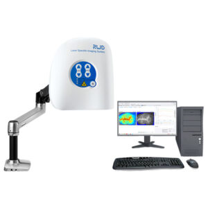
RFLSI-ZW Laser Speckle Contrast Imaging System
RFLSI-ZW laser speckle imaging system is an even better tool for microcirculation research based on laser speckle contrast imaging technology (LSCI).
With the advanced optical design and improved image processing algorithm, RFLSI-ZW shows greater performance in imaging field size, image quality, full-field frame rate and optical resolution, and provides a powerful and efficient means for human and animal tissue microcirculation measurement.
-
Laser Speckle Imaging Systems - RWD
RFLSI-ZW Laser Speckle Contrast Imaging System
RFLSI-ZW laser speckle imaging system is an even better tool for microcirculation research based on laser speckle contrast imaging technology (LSCI).
With the advanced optical design and improved image processing algorithm, RFLSI-ZW shows greater performance in imaging field size, image quality, full-field frame rate and optical resolution, and provides a powerful and efficient means for human and animal tissue microcirculation measurement.SKU: RFLSI Ⅲ-1
-
Laser Speckle Imaging Systems - RWD
RFLSI-ZW Laser Speckle Contrast Imaging System
RFLSI-ZW laser speckle imaging system is an even better tool for microcirculation research based on laser speckle contrast imaging technology (LSCI).
With the advanced optical design and improved image processing algorithm, RFLSI-ZW shows greater performance in imaging field size, image quality, full-field frame rate and optical resolution, and provides a powerful and efficient means for human and animal tissue microcirculation measurement.